State-of-the-art molecular cytogenetic techniques. The major referral laboratory in Israel both for clinical and research purposed.
FISH detects structural and numerical chromosomal alterations and provides important information of diagnostic and prognostic relevance both in hematological malignancies and solid tumors.
SKY determines the unique spectral profile of each chromosome generated by specific combinations of different fluorochromes. (Fig.1). The highly important input of SKY analysis to clinical research of malignant diseases is the identification of novel recurrent aberrations with pathogenic potential and the prediction of a specific phenotype or modified response to treatment or outcome. Combined simultaneous morphological and FISH multiparametric analyses (Fig.2) on the level of the single cell allows detection of small population of pathological cells and thus improve the accuracy of minimal residual disease (MRD) testing.
Professional scientist: Luba Trakhtenbrot PhD

DNA Sequencing
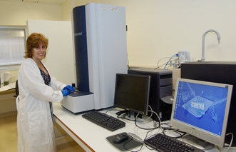
Sequenom MassARRAY MALDI-TOF MS system is a high-performance DNA analysis platform at a single base pair level resolution that efficiently and precisely measures the amount of genetic target material and variations therein. This versatile system has multi-application abilities and can be used for genotyping, methylation and quantitative gene expression analysis. In our lab we have developed a novel method for quantification A-I RNA editing levels using the Sequenom MALDI-TOF assay. We are using this system for both basic research and clinical diagnosis
Professional scientist: Michal Safran PhD
Microarray Laboratory
Affymetrix oligonucleotide based microarrays which enable full gene expression exploration for a wide range of organisms and tissues.
-
Planning and carrying out microarray experiments
-
Expression analysis services that include RNA/mRNA extraction, preparation of target samples suitable for array hybridization using both gene and all exon arrays
-
Thorough data analysis including full gene function exploration, bioinformatics and pathways discovery
-
Single nucleotide polymorphism (SNP) analysis using both Affymetrix (10,100 and 500 K assays), and Tiling arrays analysis.
-
MicroRNA arrays (will be soon incorporated)
T Functional Genomics; Global gene Expression Website
Professional scientist: Jasmine Jacob-Hirsch Msc
Zebrafish - Genetic, Developmental and Behavioral Model
The zebrafish (Danio rerio) is a powerful model organism that exhibits resemblance to human development and physiology and homology with many human genes. We are creating and studying variety of zebrafish genetic models for human diseases such as depression, developmental disorders and cancer.
Zebrafish uniquely combine features, such as small size, large clutch size, fast ex utero development and distinct behaviors, which are important for facilitating large scale studies such as drugs screening and finding the genetic source of certain diseases.
Professional scientist: Limor Ziv-Strasser, PhD
Staff: Paz Nurit, PhD student, Maayan Zoosman, Msc student
Advanced microscopy unit
The advanced microscopy unit includes several advanced microscopy-based technologies:
-
Laser capture microdissection (LCM) microscope. LCM is a method for isolating selected specific cells of interest from microscopic regions of tissue. It is a useful method of collecting selected cells for DNA, RNA and/or protein analyses thus studying changes in specific cells within a tissue. Using the LCM microscope the cells of choice are collected into a tube by activating a laser beam which catapults the cells of choice, leaving unwanted cells behind.
-
Semi-confocal fluorescence microscope equipped with a Hamamatsu ORCA-ER CCD camera. This imaging system allows the generation of confocal-like images by using the OptiGrid technology that converts the illumination system of the microscope to a structured light illumination system. The cooled color CCD Hamamatsu camera has high quantum efficiency which makes this system optimal for fluorescence/clear visual field observations of fast processes in living cells and multi-color observations.
-
Confocal Scanning laser microscope (CLSM) coupled to a multi-photon laser and a dispersion compensation unit (Zeiss LSM510 META & MaiTai DeepSee). The CLSM is a technique for obtaining high-resolution optical images. The key feature of confocal microscopy is its ability to exclude out of-focus signal from images of thick specimens. Images are acquired point-by-point and reconstructed with a computer, allowing three-dimensional reconstructions of topologically-complex objects. Our CLSM system allows spectral imaging. In addition the system is equipped with a multi-photon laser and a dispersion compensation unit. These increase the penetration into the tissue and enable imaging of live animals.
Professional scientist: Sharon Moshkovitz, PhD
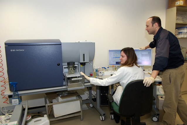
Flow cytometry and cell sorting
Polychromatic flow cytometry and cell sorting allow physical separation of specific sub-populations of cells within a heterogeneous population, with a high degree of purity, one cell at a time. Polychromatic flow cytometry is inherently capable of measuring multiple independent parameters in individual cells. For example, FACS allows monitoring the levels of intracellular phosphoproteins and transcription factors that initiate and regulate many cellular processes. Thus, for example, FACS can serve as a unique platform to specifically map in patients the signal-transduction mechanisms within cancer cells mixed among normal cells. Such profiling can be used to (1) understand signaling mechanisms that specifically characterize malignant cells, (2) describe cancers by molecular phenotype, and importantly (3) translate this information for clinical and therapeutic applications, for example to follow the effect of drugs on the phosphorylation of disease relevant kinases and/or their targets.
Professional scientist: Itamar Goldstein MD
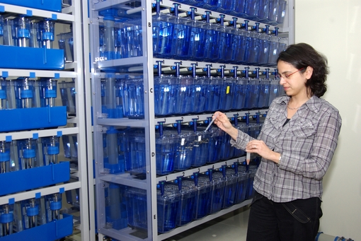
Institutional Tissue Bank - Sheba Medical Center
The Institutional Tissue Bank (ITB) at Sheba Medical Center provides researchers with high-quality and well-annotated biospecimens, essential for accelerating biomedical research and medical product development. Identification of specific markers and pathways in cancerous specimens may lead to better understanding of the biology of cancer, to the development of specific relevant therapeutics and to the establishment of the 'Translational Research & Personalized Medicine Program'.
The infrastructure of the ITB enables the collection and preservation of human tissues from patients with cancer and other diseases. Following Institutional Review Board (IRB) approval and signing an Informed Consent Form (ICF), the specimens are collected by a designated pathologist, at the operating room. Tumor and tumor-free specimens are collected, only when there is sufficient amount of tumor, in excess of the routine needs. Blood samples are collected prior to surgery/procedure. Specimens are processed immediately (10-30 minutes for tissues and up to a few hours for blood) and preserved in small aliquots. Clinical/pathological/follow-up data is collected in a designated database.
The Sheba-ITB provides the platform for collaboration. Since activation (15 months ago), we have obtained hundreds of biosamples, from different sources (thoracic, breast, brain, kidney, colon/rectum, pancreas, liver, stomach, bladder, gynecology, orthopedics, children, head & neck). These biosamples are soon to be used in research for the development of targeted therapy', facilitating pharmacogenetic studies.
All ITB activities are according to well established & validated procedures.