CT Arthrography
The purpose of CT arthrography is to demonstrate intra-articular elements. Using this examination, different joints can be imaged, including the shoulder, the hip and the ankle.
The examination is performed in two steps:
-
Arthrography: During the arthrography step, contrast material is injected into the joint by a physician. The examination is performed under fluoroscopy guidance while the patient is in the supine position. Local anesthesia is first introduced into the examined area. Then the physician advances a very fine needle into the joint and injects contrast material to fill the joint space. This procedure is generally painless but somewhat uncomfortable. Duration is generally 15-30 minutes.
-
CT examination: Computerized Tomography (CT) is a non invasive and painless imaging modality in which an image is created by projecting a controlled x-ray beam through an object (e.g. the shoulder) and extrapolating the amount of beam attenuation into a gray scale image. The data is then used to create an image of the organ examined.
A CT scan usually takes several minutes.
Preparations for CT arthrography:
Generally no special preparation is required. However if you regularly take medication or suffer from any disease please notify us when scheduling an appointment and alert the physician performing the examination.
In rare cases, the contrast material injected into the joint may induce an allergic reaction. Patients with known allergies to medications or to iodinated contrast agents and patients with known active asthma should notify their physician when scheduling an appointment in order to receive specific medical preparation.

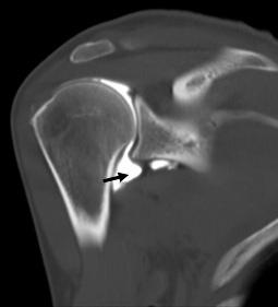
An image from a CT arthrography of the right shoulder. The arrow points to the white fluid (contrast material) injected into the shoulder joint. |
|---|
Following the examination:
During the same day you should abstain from physical activity of the examined organ. Some pain can develop when the local anesthesia wears off and may last a day or two. Pain medication can be used to relieve the pain.


