What is Cardiac CT Angiography
Cardiac CT often referred to as "Virtual coronary catheterization" is a CT scan done using a 64 slice CT scanner, in synchronization with the cardiac contraction (ECG Gating). In the Sheba Medical Center two 64 slice scanners are used for dedicated cardiac CT scanning
A Few Words regarding coronary artery disease
-
The heart oxygen demand is supplied by three major arteries: the coronary arteries (left, right and circumflex).
-
When an obstruction occurs in any of these arteries (the cause being coronary artery disease), the patient presents with chest pain , known as "angina pectoris". In case of severe coronary stenosis (obstruction), myocardial infarction follows and the patient is in great danger.
-
Preventive measures can help stop disease progression, prevent the myocardial infarction and save the patient's life.
-
Cardiac CT is a non invasive test, (not including catheter insertion into the patient's body) allowing the precise demonstration of the coronary artery walls. Significant obstruction as well as minor changes (that so minimal, they are not seen on coronary catheterization) can be demonstrated easily. The scan is very short and lasts only several minutes.
-
Many reports in the medical literature assure the high negative predictive value of the scan. That is - in the presence of either normal scan or a scan without significant obstruction the possibility of significant coronary artery disease necessitating catheterization is minimal to none.
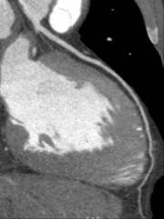
An example of a normal study
The left anterior descending artery (LAD) is seen clearly with no evidence of atherosclerotic disease, or significant narrowing
A clinical example in which Cardiac CT can save a patient's life.
A 60-year-old male with chest pain, which had improved during his stay in the emergency room. The patient was hospitalized and underwent Cardiac CT the next day.
|
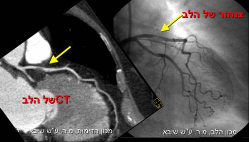
A soft plaque (arrow) can be seen in the left anterior descending artery (LAD) , causing critical narrowing (CT image on the right , coronary catheterization image on the left) resulting in chest pain during exercise (angina pectoris)
|
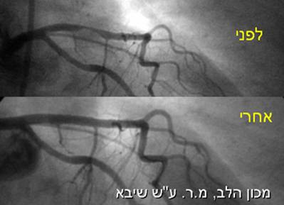
Following these findings seen on the cardiac CT the patient underwent coronary catheterization, which confirmed the finding.
The patient was treated by balloon dilatation and stent insertion in order to dilate the narrowed area preventing myocardial damage and most probably saving this patient's life
(lower image before and after treatment ).
Cardiac CT for whom is it suited
-
Patients suffering from chest pain in the presence of equivocal other non invasive test (echocardiography, cardiac nuclear medicine), or a patient that cannot perform a stress test.
-
Patients that underwent coronary catheterization and have unsolved questions/
-
Patients that underwent coronary bypass surgery, still complain of chest pain and there is a need to visualize the coronary arteries and the grafs.
-
Patients that had a coronary stent placed in a coronary artery, and there a need to verify the stent is open.
-
Patients with congenital heart defects.
-
Patients planed to undergo valve replacement, cardiac CT can visualize the coronary arteries, obviating the need for coronary catheterization.
-
Patients with an artificial valve suspected of malfunction.
-
Patients with risk factors including family history of coronary artery disease, hypertension, hypercholesterolemia, in the need to evaluate the coronary arteries in accordance with the referring physician's request.
|
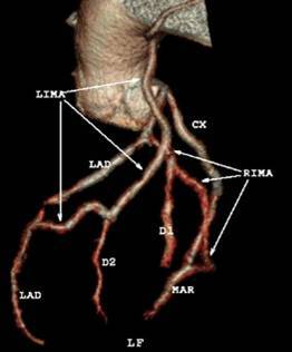
Cardiac CT in a patient following bypass surgery, all grafts are open. The scan is noninvasive simple, short, and not painful
|
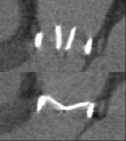
Cardiac CT can demonstrate the function of mechanical valves. Upper image: open mechanical aortic valve. Lower image: closed mechanical aortic valve. The scan is fast and reliable (only a 10 seconds scan).
|
Appropriate criteria for the use of cardiac CT were published in a consensus paper from the leading groups in Radiology and Cardiology Appropriate criteria.
How is the study preformed
-
At the Sheba medical Center Radiologists and cardiologists collaborate in order to give a high quality interpretation of cardiac CT.
-
When you arrive you will be referred to the rehabilitation center in the heart institute for ECG tracing performance. Following that you will be interviewed by a cardiologist in order to verify that the cardiac CT is the appropriate study for you, as a patient.
-
Patients who do not fulfill the appropriate criteria will not undergo the scan.
-
A medication (beta blocker) to slow the patient's heart rate might be administered in order to assure low heart rate (important for study quality). It can take up to an hour for this medication to have it's full effect.
-
Following that the patient will arrive at the Radiology department. The scan is conducted by a dedicated expert radiologist .
-
Two scans are acquired :
- the first, using a low dose setting includes scanning of the heart with no contrast injection. At the end of this scan we evaluate the scan for the presence of severe coronary calcification (which do not allow appropriate scan interpretation; in these cases the study will stop here). The first step includes scanning of the lungs, this is specifically important for chronic smokers.
- The second study includes contrast intravenous administration. The patient is required to hold his/her breath for 10 seconds.
-
The study does not cause pain to the patient, there no need for hospitalization.
-
The study will be interpreted jointly by an experienced team of radiologist and cardiologist with vast experience in cardiac imaging.
-
A summary session will be appointed within one week of the scan. The cardiologist will overviwer the results of the scan with the patient and explain it's meaning to the patient.
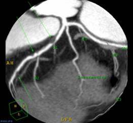
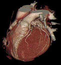
- A three dimensional reconstruction of the heart demonstrating significant (stenosis) obstructions in the left anterior descending (LAD) artery.
- New!!! Lowering the radiation exposure to 70 % in cardiac CT scanning, at Sheba Medical Center.