

MRI of the Musculoskeletal System
What is MRI (magnetic resonance imaging) of the Musculoskeletal System?
Magnetic resonance Imaging is a non invasive, essentially painless, imaging study which provides an excellent tool in evaluating a wide range of medical conditions.
MR utilizes a very strong static magnetic field, many times stronger than that of the earth's field, as well as radio waves and computer software in order to create a very detailed image of an organ, including the adjacent soft tissues, bones and bone marrow and essentially any material which contains protons. The images are created without the use of ionizing radiation.
MR allows physicians to better evaluate the bones and muscles for disease processes that may not be assessed adequately with other imaging modalities.

MRI of the knee - sagittal image The arrows point to the external (lateral) meniscus |
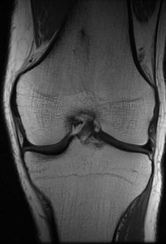
Images - MRI of the knee - coronal (frontal) image |
|---|
Equipment
- 2 state-of-the-art MR scanners (GE Healthcare)
- including one 1.5-Tesla scanner
- a new powerful 3-Tesla scanner
The stronger magnetic field provides better resolution without prolonging scan time, and also allows cutting down in the amount of contrast media without compromising image quality.
Common uses
MRI of is the modality of choice in demonstrating:
-
Joints and intra-articular elements (like the menisci of the knee)
-
The spine and disc pathologies
-
Soft tissues
How is the scan performed?
Scans are usually arranged through the Healthcare system ("Kupot Holim").
For this type of study you will be asked to lie on the examination table and be positioned within the scanner opening. The technician will then place a specially designed coil over the part of the body to be investigated. Elastic straps and pillows will help you stay in the correct position. The technician will then advance the bed into the scanner itself which is shaped like a long hollow tube and will leave the room. During the exam the scanner will make very loud rapping noises (like a jackhammer) which will require you to wear ear plugs.
A routine MR includes a series of sequences, each lasting a few minutes and may last between 30 and 60 minutes. It is important to hold still as much as possible during the scan in order to avoid blurring of the images.
For some patients, a small intravenous catheter will be placed in their arm to administer a paramagnetic contrast agent, Gadolinium. Please alert the physician or technician if you have ever experienced an allergic reaction to contrast.
Are there contraindications to MRI?
Due to the strong magnetic field in the room, it is contraindicated to undergo the study if you have certain metal hardware in your body (i.e. a pacemaker, defibrillator wires, metal implants, cochlear implants, surgical clips in the brain). Pregnant women should not have this exam unless the potential benefit from the MRI is assumed to outweigh the potential risks. In that case, you will be tested in the lower field magnet (1.5 Tesla) and contrast will usually not be administered. Some claustrophobic patients may require pre-medication to relax during the study.
Advantages:
-
The MR scan is an ideal tool for demonstrating the skeletal system with excellent ability to distinguish between different tissues including tendons and muscles that are poorly visible on plain radiographs or CT scans.
-
There is no ionizing radiation involved.
Drawbacks:
-
Long scanning times
-
Uncomfortable position and loud noises
-
Young children require general anesthesia