

MR arthrography
The purpose of MR arthrography is to demonstrate intra-articular elements. Using this examination, different joints can be imaged including the shoulder, the hip and the ankle.
The examination is performed in two steps:
-
Arthrography: In the arthrography step, contrast material is injected into the joint by a physician. The examination is performed under fluoroscopic guidance with the patient in the supine position.
First, local anesthesia is introduced into the examined area. Then the physician will advance a very fine needle into the joint and inject contrast material to fill the joint space.
This procedure is generally painless but somewhat uncomfortable. Duration is generally 15-30 minutes.
-
MR examination: Magnetic resonance Imaging is an advanced, non invasive, essentially painless imaging tool which allows a detailed assessment of a wide range of medical conditions. MR utilizes a very strong static magnetic field as well as radio waves and computer software in order to create a very detailed image of the bone, joint and the adjacent soft tissues. The images are created without the use of ionizing radiation
MRI is not painful but can be uncomfortable. An average examination lasts between 30-40 minutes.
Preparations for MR arthrography:
Generally no special preparation is required. However, if you take medication on a regular basis or suffer from any disease, please notify us when scheduling an appointment and alert the physician performing the examination.
The contrast material injected into the joint may induce allergic reactions in rare cases. Patients with known allergies to medications or to MR agents (Gadolinium) should notify their physician when scheduling an appointment in order to receive specific medical preparation.
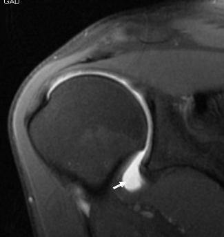
An image from MR arthrography examination of the shoulder. The arrow points to the white fluid (contrast material) injected into the shoulder joint. |
|---|
Following the examination:
During the same day you should avoid strenuous physical activity of the examined organ. Some pain can develop when the local anesthesia wears off and can last for several days. Pain medication can be used to relieve the pain.