

Conventional Radiographs (skeletal system)
What is a conventional radiograph (X-ray) of the skeletal system?
X-ray is a painless imaging modality that allows the physician to diagnose certain medical conditions and treat them accordingly.
X-ray imaging involves exposing the patient to a small amount of ionizing radiation in order to create an image.
Radiography is the oldest imaging modality but is still the most common modality in use. Basically every bone in the body may be imaged by plain radiographs including the bones of the wrist, arm, ankle, spine, etc.

An X-ray image of the wrist. The arrow is pointing to a fracture of one of the wrist bones - the Navicular, also known as the Scaphoid bone. |
|---|
Common uses for bone radiographs
-
Demonstration of a bone fracture or joint dislocation
-
Determining fracture position, stability and recovery process
-
Demonstration of joint fluid
-
Assessment prior to surgical intervention
-
Evaluation of bone involvement in infection, inflammatory diseases or growth disturbances
-
Identification and characterization of bone tumors
-
Foreign body identification (e.g. metal fragments)
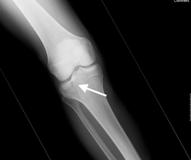
A frontal X-ray of the knee The arrow is pointing to a fracture in the tibia |
|---|
How is the X-ray performed?
The Technician will position you on the imaging table with an X-ray film or digital cassette under the table, below the imaged organ. You will be asked to avoid any movement in order to allow for a sharp and clear image.
Bone radiography usually includes two views of the imaged organ, thus the technician will adjust the position of the imaging part for a second radiograph.
Sometimes imaging of the other extremity is necessary for comparison.
Imaging may take between 5 to 10 minutes