Computerized Tomography (CT) is a non invasive and painless imaging modality in which an image is created by passing a controlled x-ray beam through an object (e.g the skull) and extrapolating the amount of beam attenuation into a gray scale image.
CT images represent the differences in tissue density (in simple terms, the denser the object the "brighter" it is on CT e.g metal, bone, calcifications and contrast; whereas fluid, fat and air which are less dense will appear "darker"). In certain instances there is a need for I.V administration of iodine based contrast material in order to accentuate the differences between the tissues. It is of most importance that the patient notify the physician or technician of any known allergic reaction to iodine or other contrast material as well as other pre-morbid conditions such as asthma, diabetes, kidney disease and thyroid related disease.
You may be asked to refrain from food and drink for 4 hours prior to the exam.
Women of child bearing age should notify the physician of any chance of pregnancy, since CT carries the risks of ionizing radiation to the fetus.
Women who are breast feeding should refrain from feeding up to 24 hours following the exam.
During the exam the patient is required to lie still on the examination table which will advance in to the rounded doughnut shaped gantry. the gantry houses the x-ray tube which will rotate in a circle around the patients head. Most studies last between 2 and 7 minutes. The images are digitally transferred via our Picture Archiving and Communication System (PACS), to dedicated workstations in which the radiologist can view them on screen.
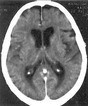
CT scan allows us to
Demonstrate the brain's anatomy, shape and size.
Identify and classify intracranial processes such as space occupying lesions- tumors, hemorrhages, abscess, infectious processes and infarcts.
Detect skull and facial bone fractures
Portray internal injuries and intracranial bleeding in a timely manner in urgent cases which allows for immediate intervention.
CT Angiogram, which requires intravenous injection of contrast, gives an accurate road map of both the extra- and intracranial vessels in search for occlusion, dissection, aneurysm formation and other vessel wall abnormalities.
CT is less sensitive than MRI in demonstrating fine abnormalities in brain texture. It has a very limited role in detecting meningitis.
To the Hebrew web site press here