

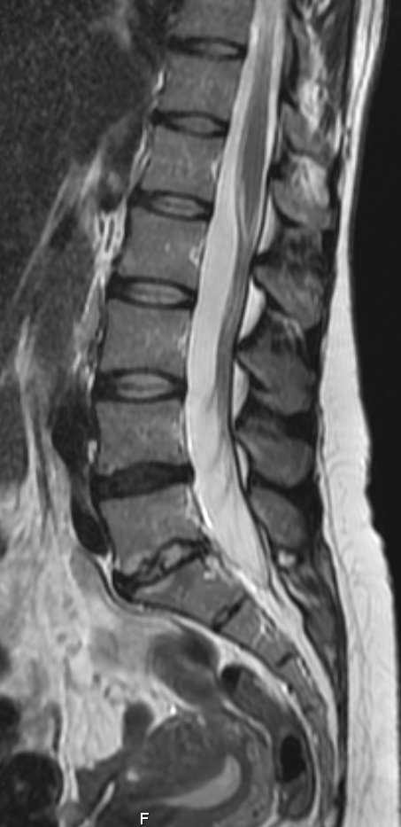
MRI OF The spine
Magnetic resonance Imaging is an advanced, non invasive, essentially painless imaging tool which allows a detailed assessment of a wide range of medical conditions. It allows physicians to evaluate the spine and spinal canal for disease processes that cannot be assessed adequately with other imaging modalities.
How does it work?
MR utilizes a very strong static magnetic field (many times stronger than the earth's), as well as radio waves and computer software in order to create a very detailed image of the spinal cord, nerve roots, adjacent soft tissues, and bone marrow. The images are created without the use of ionizing radiation.
Sheba Medical Center uses 2 state-of-the-art MR scanners (by GE Healthcare), including one 1.5-Tesla scanner and a new powerful 3-Tesla scanner. The stronger magnetic field provides better resolution without prolonging scan time and also allows cutting down in contrast material without compromising image quality.
Common uses for MR of the spine:
MR is the modality of choice in the following conditions:
- Spinal cord injury
- Intervertebral disc bulges/protrusions
- Preoperative planning
- Congenital anomalies of the spine
- Infectious processes of the vertebra or cord
- tumors
MR is also frequently performed for investigation of:
· Tumors - diagnosis and follow up
· Neck pain and suspected disc protrusion
· Lower back pain and suspected disc protrusion
· Backache accompanied with fever to rule out infection
· Upper or lower limb parasthesias suspected cord injury
· Fetal imaging when ultrasound is inconclusive
How is the scan done?
Scans are usually ordered through the Healthcare systems ("Kupot Holim").
For this type of study you will be asked to lie on the patient table and be placed within the scanner opening. Then the technician will place a specially designed coil over the part of the body to be investigated. Elastic straps and pillows will help you remain in the correct position. The technician will then advance the bed into the scanner itself which is shaped like a long hollow tube and will leave the room. During the exam the scanner will make very loud rapping noises (like a jackhammer) which will require you to wear earplugs.
A routine MR scan of the head may last between 30 and 60 minutes and includes a series of sequences, each lasting a few minutes. It is important to hold still as much as possible during the scan in order to avoid image degradation.
For some patients, a small intravenous catheter will be placed in the arm to administer a paramagnetic contrast agent, gadolinium. If you ever had an allergic reaction to gadolinium, please alert the physician or technician.
Are there contraindications?
Due to the strong magnetic field in the room, it is contraindicated to undergo the study if you have certain metal hardware in your body (i.e. a pacemaker, defibrillator wires, metal implants, cochlear implants, surgical clips in the brain). Pregnant women should not have this exam unless the potential benefit from the MRI is assumed to outweigh the potential risks. In that case, you will be tested in the lower field (1.5 Tesla) magnet and contrast will usually not be administered. For claustrophobic patients, some may require pre-medication to relax during the study.
Advantages:
· The MR scan is an ideal tool for demonstrating marrow signal in the vertebrae and spinal cord substance with superb ability to discriminate between the tissues.
· No artifacts from surrounding bony structures and shoulder girdle
· no ionizing radiation involved.
Drawbacks:
· Long scanning times
· Uncomfortable position and loud noises
· Young children require general anesthesia