What is a CT?
Computerized tomography (CT) is painless, noninvasive method of diagnosing and treating a wide variety of diseases and conditions.
CT uses an x-ray beam and a high speed computer to create images of cross sections of the body which can be viewed on a computer or on film. This type of imaging gives precise, detailed information on tissues of the body, distinguishing them by their density. It is much more sensitive than a regular radiographic film.
During the exam we are careful to minimize the radiation exposure to each child according to the standards of the American Society for Pediatric Radiology.
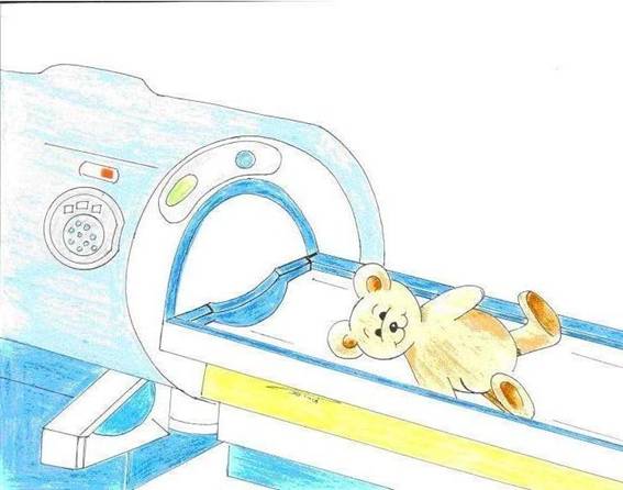
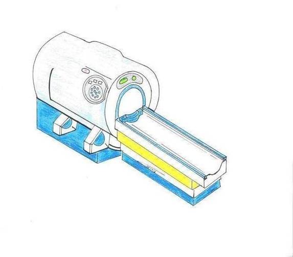
What is the appearance of the CT machine?
The CT machine is a large, doughnut shaped device with a sliding table within the hole, or gantry. The X-ray "tube," the radiation source, and the detectors rotate around the patient passing through the gantry on the table. Generally speaking images are obtained in the axial plane (like salami slices), but can be reconstructed in any plane, using the same information obtained during the examination without additional radiation exposure.
What takes place during the exam?
During the exam your child will lie on the table and a technologist will position him properly for the exam. The child will pass through the gantry several times during the exam, each time for several seconds.
In the event of intravenous contrast injection, your child will feel a small prick as the catheter is placed into a vein under the skin. During the injection, there is often a feeling of slight warmth all over the body and occasionally a metallic taste in the mouth. These are short-lived, expected and normal occurrences. For abdominal imaging, oral contrast material is drunk. This liquid is flavored with syrup to mask a slightly bitter taste. Most children are able to drink this contrast with some encouragement.
Infants and small children are usually not able to cooperate by holding still, even for the short period of the exam. For this group of patients (usually up to age 6) we perform the exam under sedation.
What exams are performed in CT
Head including brain, orbits and ears; neck; chest; abdomen and pelvis; spine, joints and extremities; angiography (blood vessels).
|
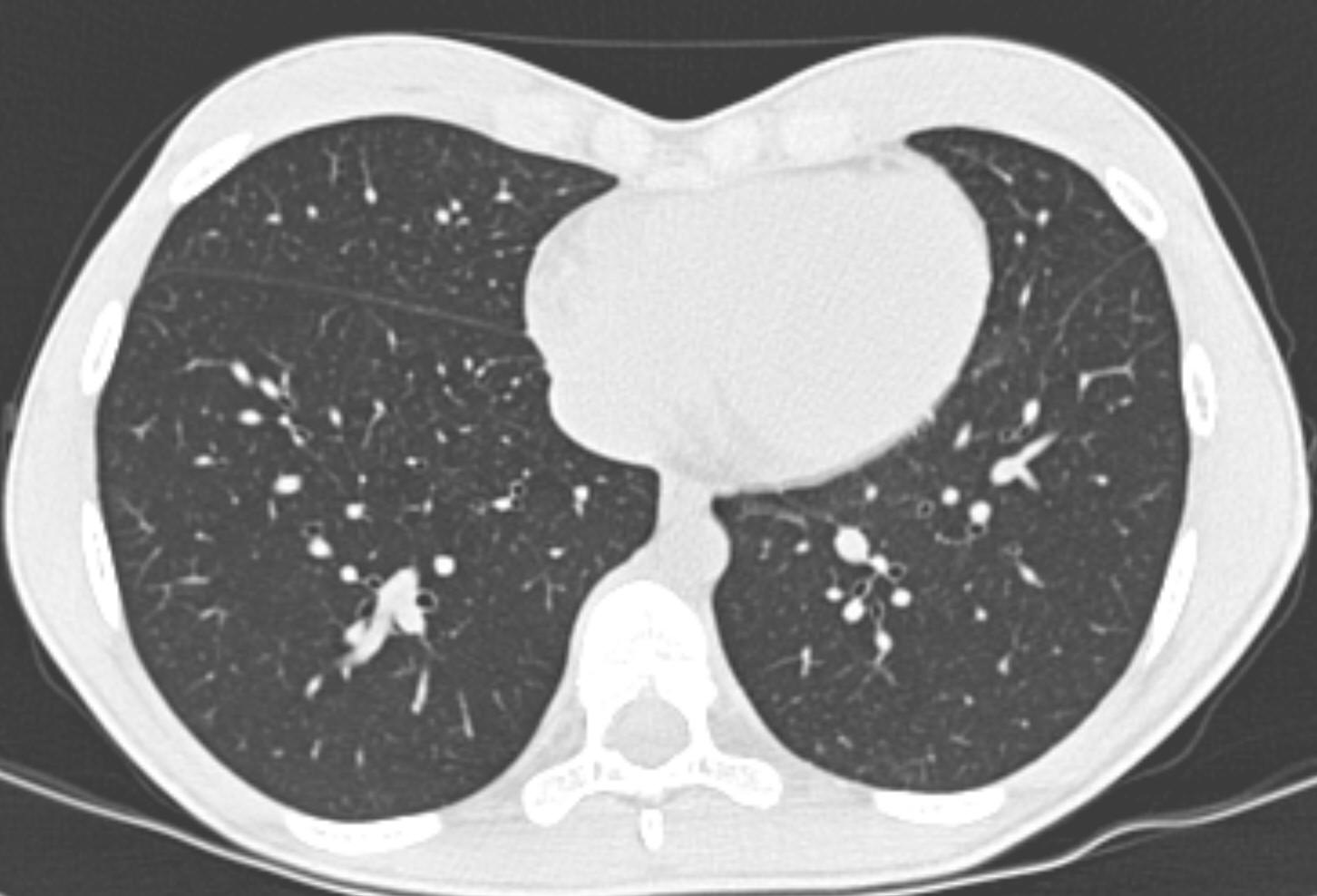
A transverse (axial) "slice" from a lung CT
|
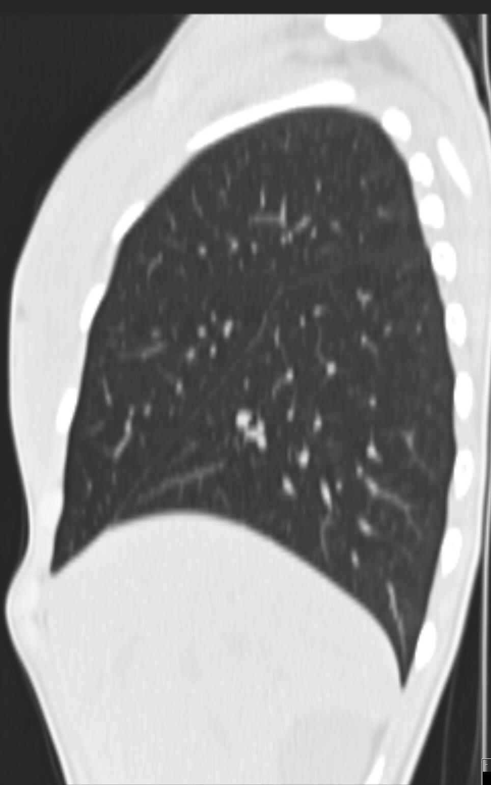 |
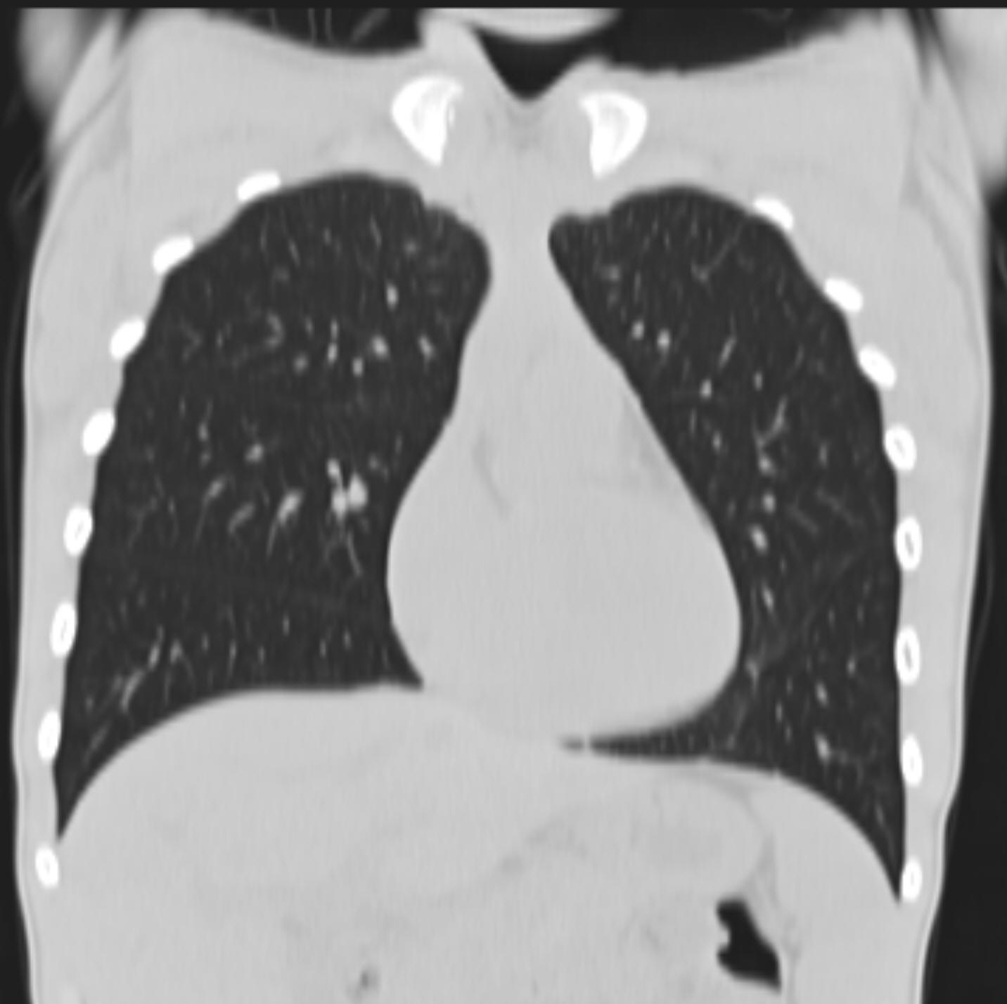 |
| Slices obtained by reconstructing information on a chest CT in the frontal (coronal) and side (parasagittal) views |
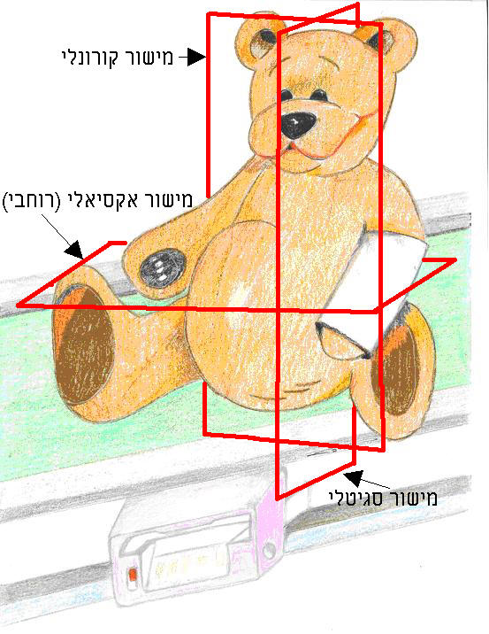
Example of the different planes one can reconstruct CT data
|