At Sheba Medical Center advanced Magnetic Resonance Imaging (MRI) is performed on fetuses, infants and children. MRI is a diagnostic tool requiring no ionizing radiation, a great advantage when performing imaging studies on children. MRI is based on strong magnetic fields and radio waves. They are utilized to create images with excellent tissue characterization, in multiple viewing planes and in minute detail. MRI on children is performed under the guidance and supervision of pediatric imaging staff physicians in the MRI center. This section of the main imaging department is located at the base of the main inpatient tower.
In young children (usually less than 5 yrs, but possibly any child) this exam is performed under general anesthesia. This is due to the relatively long exam time (30-60 minutes) and the sensitivity of this type of imaging to motion. During the exam the patient has to lay almost motionless for long periods of time. The MRI machine (or magnet) makes thumping noises during imaging.
Occasionally, for a portion of the test, the older child will be ask to hold his breath for a few short seconds to prevent even respiratory motion during image acquisition. The parent can be in the room at the time of the imaging to touch and speak to the child in order to relax him. Of course any child who is able can have the exam performed without anesthesia.
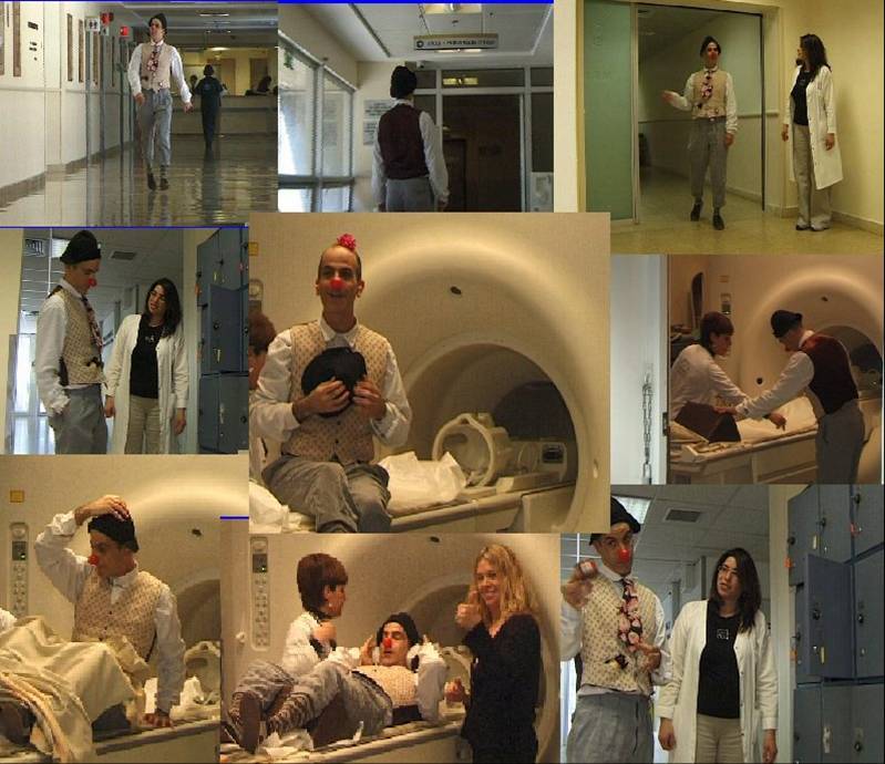
We would be happy to familiarize any child scheduled for an exam to have a tour of the facility several days before the exam and answer any questions he may have in order to be able to maximize his cooperation during the exam.
MRI studies performed at Sheba Medical Center
Chest, Abdominal and Pelvic MRI- These are often performed on oncology patients, but also to answer tissue specific questions and on patient's who are allergic to contrast or cannot have even relatively small doses of ionizing radiation.
MRCP
MRCP Magnetic Resonance Cholangiopancreatogram
MR examination of the bile ducts and pancreatic ducts performed in two and three dimensions usually performed to investigate congenital abnormalities of the biliary tree, long standing jaundice and pancreatitis.
MRE Magnetic Resonance Enterography
Advanced diagnostic imaging of the intestines usually in work up or follow up of patients with inflammatory bowel disease.
MRU Magnetic Resonance Urography
A relatively new exam designed to provide precise anatomic and functional information on the urinary tract. In one single test it provides most if not all information provided by ultrasound, pyleography, cystourethrography and nuclear scintigraphy. This cutting edge study includes basic images acquired before and during excretion of contrast and analysis of data which is performed later on a separate specially designed work station
Fetal MRI
This examination is performed for the most part after the 23rd week of pregnancy. No harmful effects on an unborn child from an MRI exam have been reported in the literature. These exams are performed without sedation and without injecting contrast into the mother. They are usually requested for further evaluation of findings found on obstetrical ultrasound examination.
This study provides both 2D and 3D images of the entire fetus allowing for further evaluation of masses, cranial anomalies, diaphragmatic hernia and anomalies of the urinary tract and digestive system. The results of the study are interpreted in a timely manner and discussed with a team of doctors including pediatricians, geneticists, obstetricians and radiologists.
MRA/MRV Magnetic Resonance Imaging of the Vascular System
This examination is performed for evaluation of the vascular system and the heart in patients with congenital or developmental anomalies or abnormalities acquired through disease, surgery or trauma. It may be performed with or without contrast injection depending on the disease the query of the requesting physician.
MRI of the Pediatric Central Nervous System
These exams are performed at Sheba Medical Center by the Neuroradiology section of the Department of Diagnostic Imaging. Dr. Chen Hoffman oversees the performance of these tests as well as MRI of the fetal central nervous system.
Cardiac MRI
These examinations are performed in children in conjunction with the Cardiac Imaging section under the guidance of Prof. Eli Konen, the director of the Department of Diagnostic Imaging and Dr. Goitein, both of whom are fellowship trained in cardiac imaging and have the most experience and expertise in Israel in Cardiac Imaging.
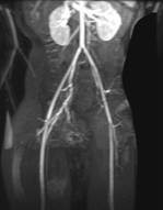
MR angiography of the vascular supply of a lymphangioma using 3D reconstructions.
|
Preparation for a Pediatric MRI examination
A detailed explanation of what to expect during an MRI exam is given to each child according to his understanding, very important for increasing the likelihood of a successful examination (Please see the included video clip).
The child should be dressed in comfortable, light clothing which do not contain metal such as snaps, buttons buckles, etc. Jewelry and other valuables should be left at home. Each person entering the examination room will be asked to fill out a questionnaire at the reception desk and submit to a metal detector test for their own safety and the safety others in the MRI suite.
Children scheduled for an MRI examination under general anesthesia should come to the imaging center after fasting at least 6 hours, including solid and liquid food. Clear fluids can be given in small amounts up to 3 hours prior to the exam. Breast feeding infants should fast at least 3 hours prior to the exam.
|
IV Contrast Injection?
|
IV Fluids Required
|
Oral Prep Required
|
Fasting
|
Time of Arrival for Exam
|
Exam
|
|
Dependent on Exam
|
Dependent on Exam
|
No
|
6 Hours
|
Hour prior to exam
|
MR under anesthesia
|
|
Yes
|
No
|
No
|
Not required
|
Time scheduled.
|
Chest/Abdomen
|
|
Yes
|
No
|
Yes
|
6 Hours
|
Hour prior
|
MRE
|
|
Yes
|
Yes
|
No
|
|
Hour prior
|
MRU
|
|
No
|
No
|
No
|
|
Time scheduled
|
MRCP
|
|
Yes
|
No
|
No
|
|
Time scheduled
|
MRA
|
|
No
|
No
|
No
|
4 Hours
|
Time scheduled
|
Fetal MRI
|