Ultrasound Examinations in Children
Ultrasound imaging utilizes sound waves with frequencies that are not heard by the human ear. These waves enter the body through a transducer and are either absorbed, reflected or transmitted. The reflected waves, or echoes are used to depict an images of the internal organs in real time. This imaging modality does not use ionizing radiation and therefore is an ideal imaging tool in the fetus, infant and young child and adolescent, whose growing bodies are more sensitive to radiation damage.
In general there is no special preparation for most ultrasound examinations.
The parent can accompany the child and, contrary to radiographic and fluoroscopic examinations, even a pregnant mother may stay in the room during the examination.
The main structures examined by ultrasound are the abdominal organs as well as certain organs in the chest and neck, the musculoskeletal system and soft tissues.
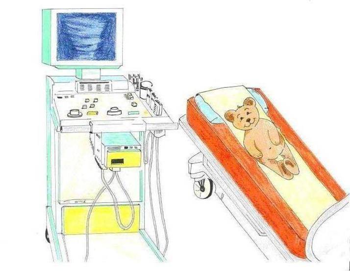
By means of a computerized imaging machine and a small wand called a transducer which is placed on the skin after applying gel, the ultrasound machine broadcasts sound waves into the body and listens for the echoes which return from the body back to the transducer. These echoes are processed based on their strength, where on the transducer and when they returned and are displayed on monitor.
Because images are displayed in real time, motion of muscles, bodily fluids and digesting material can be detected. Blood flow can be studied using color and pulse wave Doppler.
Bones, because they absorb sound, and gas, because it reflects sound, are not ideal media for ultrasound examinations. Only the outer layer, or cortex, of bone can be seen by ultrasound. Bodily structures surrounded by bones are often obscured. Gas in the digestive tract and lungs make these organs unsuitable for ultrasound examination under normal circumstances. However, fluid around or in the lungs and fluid filled bowel loops can be imaged. Gas in the bowel can be moved by compression or patient positioning.
Abdominal and Renal Ultrasound Examinations
These examinations are performed to investigate abdominal pain, abdominal abnormalities felt by the pediatrician on physical exam such as masses or enlarged organs and infections of the urinary tract, among others. They are also requested to follow up on known abnormalities such as tumors, dilatation of the urinary tract or complications after surgery.
Preparation for abdominal ultrasound: Fasting: Up to 3 years of age fast the length between meals/feedings, from 3-6 hours fast of 4 hours, over 6 years fast of 6 hours.
Preparation for urinary tract ultrasound: Under 3 years of age no preparation is necessary, Over 3 years of age voiding 2 hrs before the exam and then drinking 2-3 cups of water immediately afterwards with no further voiding until after the exam.
Spinal Ultrasound Examination:
This exam is usually performed in infants less than 4 months of age referred by neonatologists or from infant well care centers to evaluate the lower spinal cord, soft tissues of the lower back and osseous structures of the lumbar and sacral spine.
No preparation is required.
Neck, Soft Tissue and Musculoskeletal Ultrasound Examinations:
Performed to investigate congenital or acquired abnormalities referred by pediatricians to further evaluate findings on physical exam or on plain radiographs.
No preparation is required.
Head Ultrasound Examination:
Contrary to the case in adults, the brain of an infant is able to be imaged by ultrasound to the presence of the anterior fontanel or soft spot of the skull usually up to at least 8 months of age. The younger the infant the better the images will be as the brain is smaller allowing use of higher resolution frequencies and the fontanel is larger. As the fontanel closes in older infants the exam becomes more limited. The advantage of the exam is the lack of ionizing radiation in its performance, and the lack of necessity for sedation or anesthesia as needed for CT and MR.
The exam is primarily performed to screen and follow up premature infants and to evaluate abnormal head circumference measurements.
No special preparation is required.
US guided Biopsies, Aspirations and Drainage Procedures:
Histological and Cytological specimens can be obtained under US guidance.
These procedures are performed at the request of referring doctors in the Department of Pediatric Radiology.
Most of these procedures require sedation and/or light anesthesia.
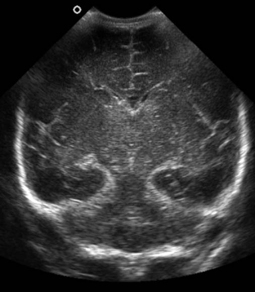
Head US examination performed through the anterior fontanel or soft spot in the skull in the coronal plane. The visualized structures are symmetric and normal.
|
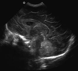
Head US examination performed through the anterior fontanel or soft spot in the skull in the sagittal plane demonstrating the normal midline structures of the brain
|